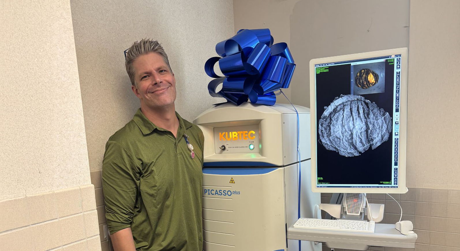
Case Studies Utilizing KUBTEC Mozart 3D Tomosynthesis for Margin Assessment during Partial Mastectomy by Andrea Madrigrano, MD
Access Case StudiesDr. Andrea Madrigrano, associate professor in the Department of Surgery at Rush University Medical Center shares her experience utilizing Mozart 3D Specimen Tomosynthesis for accurate margin assessments in her practice.
"Specimen tomosynthesis allows me to remove a smaller volume of initial incision allowing patients to have a better cosmetic result” - Andrea Madrigrano, MD
Case 1
- 80-year-old female with screen-detected LEFT breast focal asymmetry with diagnostic imaging confirming a 1 cm X 0.8 cm X 0.9cm mass
- Core biopsy- IDC, grade 1, ER 100, PR100, Her 2 1+
- Left breast lumpectomy: Invasive ductal carcinoma, grade 1, 0.6 cm
- Additional superior margin; excision: Benign breast tissue. No evidence of atypia or malignancy.
- Additional anterior margin; excision: Benign breast tissue. No evidence of atypia or malignancy.
Case 2
- 50-year-oldwith screen-detected LEFT breast microcalcifications with stereotactic biopsy showing DCIS. MRI showed no residual enhancement and marking clip 1cm inferior to the location of core biopsy.
- Final Pathology: DCIS, 2 mm, 0.5 posterior, and0.5 mm inferior margins of resection.
- Dissection carried to the fascia
- Medial Margin: Benign breast tissue
- Inferior Margin: Benign breast tissue
Case 3
- 63-year-old female found to have two small, low-grade cancers at the 3 and 6 o’clock position on screening mammogram. She had a large breast with grade 3 ptosis.
- Final Pathology: 3 o’clock IDC, grade 2, 1.2cm,with DCIS, margins clear
- 6 o’clock, IDC, grade 1, 1.5cm, 2mm from anterior and posterior margin
- Additional anterior, posterior, and inferior margin benign breast tissue
Case 4
- 66-year-old with screen-detected breast cancer
- A spiculated mass is demonstrated in the mid superior slightly lateral right breast. This has some borders which are spiculated and others which are obscured. Some faint calcifications are noted throughout this mass. The dominant mass extends over about 3 cm with possible subtle spiculations extending further away.
- Right breast; lumpectomy: Invasive ductal carcinoma, grade 3, measuring 2.5 cm in greatest dimension. Invasive tumor is present 0.5 mm from the superior margin of excision
- Ductal carcinoma in situ (DCIS) grade 3, solid type, with necrosis and associated with microcalcifications
- In situ component present at the superior margin of excision. Previous biopsy cavity changes identified.
- (Right breast, superior margin; excision) Benign breast tissue with previous biopsy cavity changes
- (Right breast, medial margin; excision) Benign breast tissue, negative for malignancy
Case 5
- 63-year-old female with large, ptotic breasts found to have extensive microcalcifications found on mammogram spanning 7cm AP,5.5 cm SI and 6cm ML. Core biopsy showed DCIS
- Left breast mass; excision: Invasive ductal carcinoma, grade 2. Four foci: The largest measuring 3mm in linear extent. Surgical margins are free.
- Ductal carcinoma in situ, nuclear grade 3,Ductal carcinoma in situ comprises greater than 50% of the entire tumor mass. Surgical margins are free
- Additional medial margin; excision: Ductal carcinoma in situ, nuclear grade 3. Surgical margins are free.
Case 6
- 47-year-old female found to have increasing microcalcifications on yearly mammogram. These spanned over a 7-8 cm area.
- Right breast, partial mastectomy: Ductal carcinoma in situ (DCIS), Grade 2
- Largest focus measures 9mm, focally extending to the inked medial margin.
- Additional medial margin inferior, excision: the microscopic focus of Ductal carcinoma in situ (DCIS), cribri from type, present3mm from the margin. The margin of resection is free. Fibro-adenomatous changes and sclerosing adenosis.
- Additional medial margin superior, excision: Benign breast tissue with associated microcalcifications
.png)
Surgeon Biography:
Dr. Andrea Madrigrano is Breast Surgeon in Chicago, IL and is affiliated with Rush University Medical Center. She received her medical degree from University of Illinois College of Medicine and is completed a Surgical Breast Fellowship at Stanford University Medical Center.
Request your personal meeting or demo
Fill out the form and one of our exhibition managers with be in touch about scheduling your personal meeting or demo at our upcoming trade show.
For more news, views, & events, please visit our LinkedIn page
Click Here


