Case Studies Utilizing KUBTEC Mozart 3D Tomosynthesis for Margin Management during Partial Mastectomy by Ian T. Greenwalt, MD
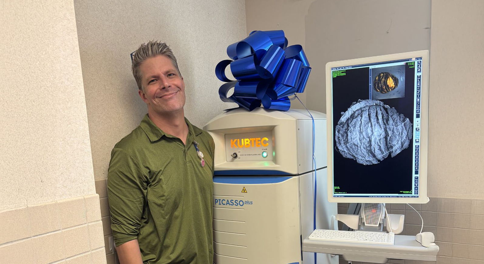
Access Case StudiesAttending surgeon at MedStar Georgetown, Ian Greenwalt, MD provides two cases where he utilized the MOZART® Specimen Tomosynthesis Imaging System from KUBTEC Medical Imaging. Dr. Greenwalt shares the operating plans for each case and how 3D tomosynthesis technology aided in real-time intraoperative decision-making.
“It gives me one more tool in the operating room to be as accurate as possible” - Dr. Ian T. Greenwalt
Case #1
• 61F postmenopausal h/o anxiety, fibromyalgia, OSA, lyme disease (well controlled, no steroids, biologics) mother h/o breast ca at 47. Previous right breast biopsy (benign). No complaints, asymptomatic, exam within normal limits.
• Bilateral sMMG – Cluster of classifications right superior breast. BI-RADS: 0-incomplete, recommended for additional imaging.
• Right dMMG - suspicious calcifications in the right breast. Biopsy recommended.BI-RADS:
• Right breast stereotactic biopsy – R DCIS, grade II-III, ER >95% PR 80-90% (hydromark barrel)
Case #2
• 64post-menopausal on HRT (insomnia, vaginal dryness) noted palpable abnormality in UOQ of L breast. Found to have 1 cm palpable mass on clinical exam and palpable left axillary lymphadenopathy.
• Left dMMG & U/S – Left UOQ, 2OC 1.2 cm mass corresponding to palpable area of concern. Left axillary lymph node with thickened cortex 1.1 cm. BIRADS 4
• Left U/S guided biopsy – Left breast 2 OC: IDC, grade III, ER (100%) PR (100%) HER2negative, Ki 67 70% LeftAxilla – Poorly differentiated carcinoma consistent with metastasis from breast primary. Staging scans negative
Discussion
• How 3D tomosynthesis provides critical information about anterior and posterior margins relative calcifications in specimen that 2D imaging may have missed.
• How 2mm margins are sufficient to understand the extend of calcifications within the specimen
• How tomosynthesis and measurement features on machine are helpful to assess inferior margin.
.png)
Surgeon Biography:
Dr. Ian T. Greenwalt is a board-certified breast surgeon practicing in the MedStar Breast Health Program at MedStar Georgetown University Hospital. A graduate of Boston College, Dr. Greenwalt received his medical degree from Drexel University College of Medicine.
Request your personal meeting or demo
Fill out the form and one of our exhibition managers with be in touch about scheduling your personal meeting or demo at our upcoming trade show.
For more news, views, & events, please visit our LinkedIn page
Click Here