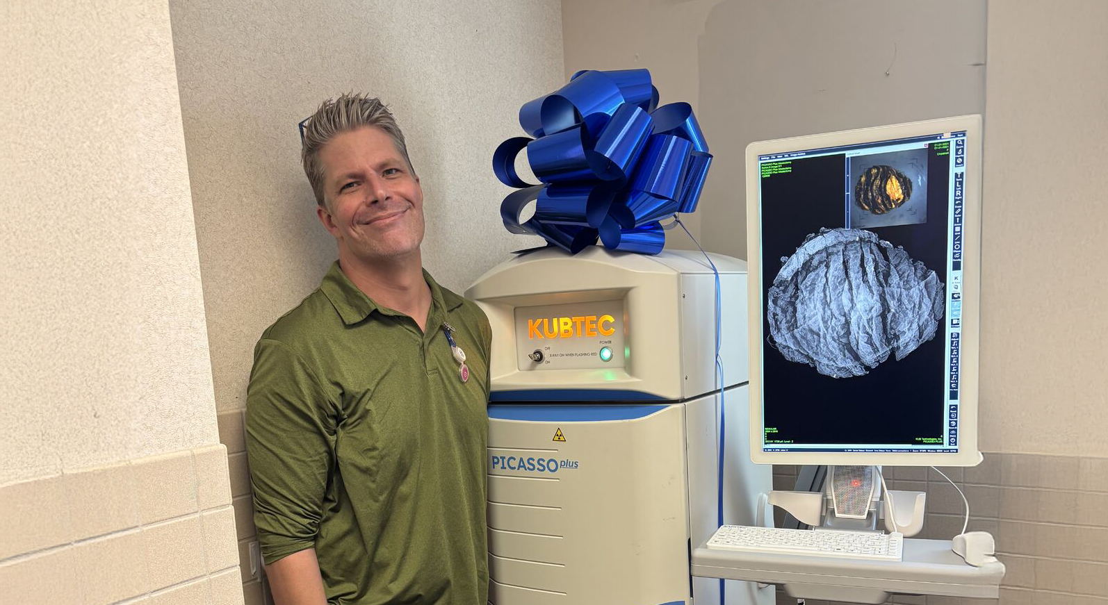
Case Studies Utilizing KUBTEC Mozart 3D Tomosynthesis for Margin Management during Partial Mastectomy by Lisa Babashoff, MD
Access Case StudiesDec 22, 2021
/
Case Studies
Dr. Lisa Babashoff, Medical Director of the Breast Surgery Program at St. John’s Regional Medical Center shares how the Mozart System has impacted her practice.
"The MOZART System has limited the amount of re-excisions that I will have to do. 3D imaging is great, it's amazing to see right there in surgery at the time it is being done” -Lisa Babashoff, MD
Case 1
- 86 y/o female with a 9mm left breast invasive ductal carcinoma, Grade 2, ER+PR+Her 2- with no family history that was picked up on screening imaging
- PMH: HTN
- She opted for lumpectomy without nodal sampling secondary to her age
Case 2
- 77 y/o female 9mm invasive ductal carcinomaER+PR+Her2- to the right breast UOQ found on screening imaging.
- Postmenopausal, DM, HTN
- Her mother was also diagnosed with breast cancer at age 77 and her daughter had ovarian cancer diagnosed at age 50. She had not had any germline mutation studies at the time
- Physical exam was negative
- She opted for lumpectomy and node sampling
- Final pathology showed a 1.2cm IDC with negative margins
- Extra shave margins from the inferior and lateral margins as dictated by the intra-op imaging that were negative.
Case 3
- 65 y/o female with two nodules noted on screening imaging in the right breast 2 and 3 o’clock position. One measured4mm and the other 5mm on imaging
- Both were biopsy-proven invasive ductal carcinoma Grade 2, ER +PR+Her2-
- Her mother was diagnosed with breast cancer at age 70
- MRI was obtained that showed 2 lesions 4 cm apart
- She wished to proceed with lumpectomy which was done through a NAC incision
Case 4
- 64 y/o female with a sister who had breast cancer diagnosed in her 40’s
- The patient has had negative mammograms most recently in April
- With her family history the patient pursued an MRI of her breast back in January 2020. This showed small areas of enhancement in both the right and left breast but more prominent in the LIQ of the left breast
- She has been undergoing 6-month MRIs since then. The most recent MRI showed 2 lesions. The first area was in the lower inner quadrant and was a 1.1cm area of non-mass enhancement. A second area in the lower outer breast was also seen that measured 2.8 cm
- Both areas showed DCIS ER+PR+ on core biopsy.
- Patient really wished for a lumpectomy which was performed through a single inferior mammary incision.
- Sentinel lymph nodes were negative, and pathology showed 7cm of DCIS with multiple positive margins
- She is to undergo re-excision in the near future
Case 5
- 66 y/o female with 1.1 cm invasive ductal carcinoma ER+PR+Her 2- in the 2 o’clock left breast
- H/o obesity
- She underwent lumpectomy and node sampling
- At the time of surgery her tumor was very lateral in the axillary tail and just superficial to the chest wall.
Lessons Learned
- Take fewer margins which saves time and expense in the operating room. This is done without leading to an increase in re-excision rates. This also leads to less potential cosmetic deformity which thereby increases patient satisfaction.
- Learned to use caution with intraoperative imaging in the DCIS patients as we know mammography does not always show the whole story.
.png)
Surgeon Biography:
Dr. Babashoff is Board Certified in General Surgery and the Medical Director of the Integrated Breast Center at St. John’s Regional Medical Center.
Request your personal meeting or demo
Fill out the form and one of our exhibition managers with be in touch about scheduling your personal meeting or demo at our upcoming trade show.
For more news, views, & events, please visit our LinkedIn page
Click Here