To ensure all cancer is removed during a surgery, the standard procedure is for the surgeon to include a border of tissue surrounding and beyond the edges of the visible cancerous tumor. This border of tissue is called the surgical margin or margin of resection, and it needs to be examined microscopically by a pathologist to determine if cancer cells are present. The pathologist measures the distance between the outer edge of the tumor containing cancer cells and the outer edge of the perceived normal tissue (the surgical margin). The results of this examination are labeled as Negative, Positive, or Clear; and each influences treatment decisions. For example, if cancer cells are still present in the surgical margin, additional surgery and radiation may be indicated. Breast cancer surgical margins are typically checked after surgical biopsy, lumpectomy, and mastectomy.
Negative Surgical Margin means that there are no cancer cells found at the edge of the tissue that was removed. In other words, the rim of normal tissue is free of cancer cells. Sometimes the distance between the cancer cells and normal tissue cells is included. It is important to note that there is no standard of how wide a “clear margin” has to be.
Positive Surgical Margin (aka Involved) means that there are cancer cells in the tissue that was perceived to be normal by the surgeon. Usually, more surgery is indicated to remove the remaining cancer.
Close Surgical Margin (aka Narrow Surgical Margin) means that the cancer cells are very close to, but do not touch, the rim of normal tissue that is free of cancer. More surgery may be indicated, depending on the type of cancer (Types of Breast Cancer | Memorial Sloan Kettering Cancer Center (mskcc.org)

After breast cancer surgery, the removed breast tissue may also be X-rayed to further conclude that the entire tumor was removed. The goal of X-raying is to see if microcalcifications that were found on a pre-surgical mammogram are not present on a post-surgical mammogram.
More about Positive Surgical Margins
When pathology indicates positive margins, chances are good that some of the tumor is still present in the breast. This situation indicates the need to obtain negative final margins (requiring another surgical procedure known as re-excision), which has significant physical, psychological, and financial implications for the patient. It means prolonged recovery, time away from work and family, and added anxiety.
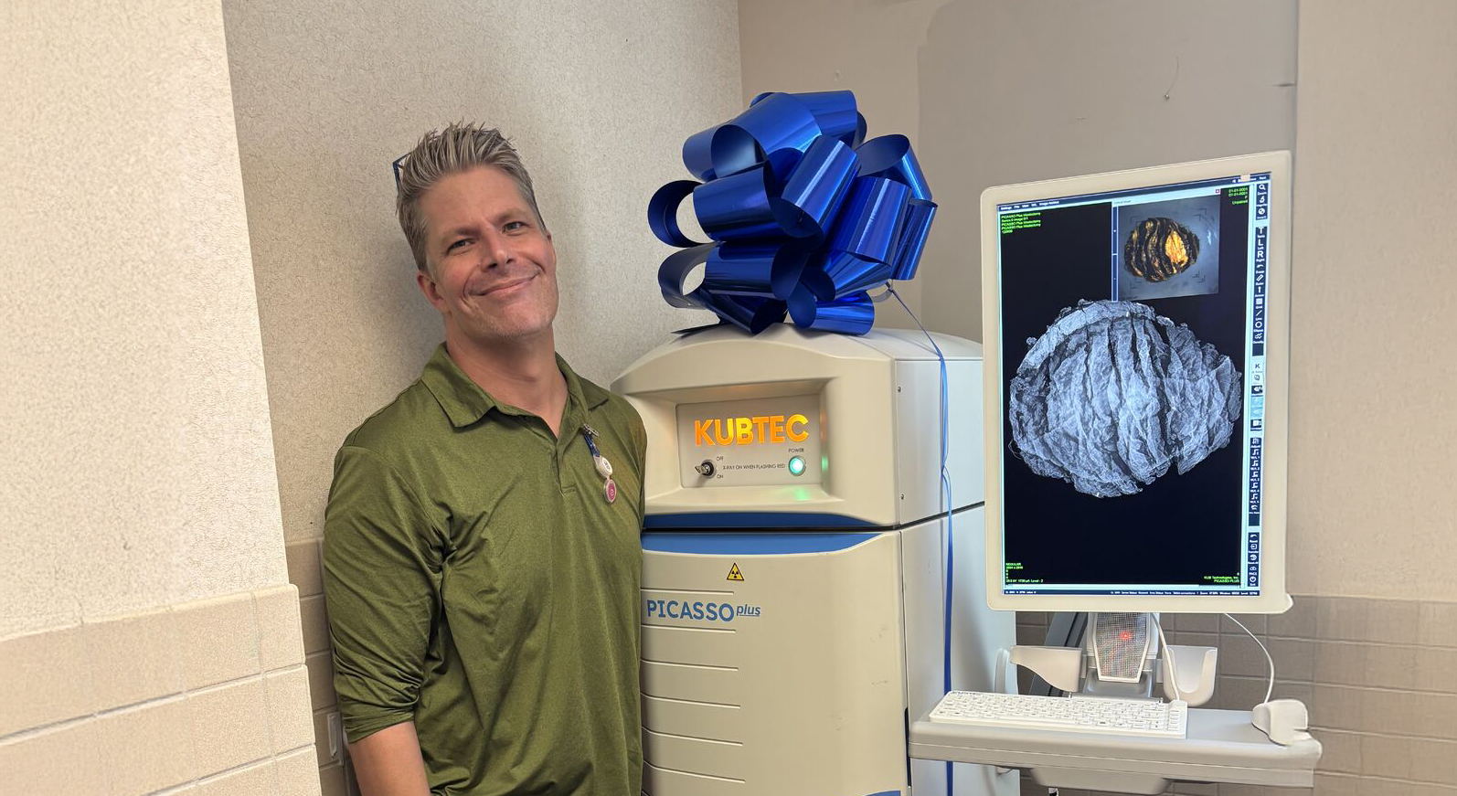
The challenge of obtaining negative final margins during the initial biopsy procedure lies in using accurate, real-time, intraoperative margin assessment techniques. One company, by the name of KUBTEC, has proven technology to do just that with their MOZART System for Specimen Tomosynthesis.
KUBTEC’s MOZART system enables analysis of the specimen in 1-millimeter digital slices. Each slice shows its own margin, and can be viewed independently of all other slices. In a traditional 2D specimen X-ray, the three-dimensional breast anatomy is compressed into a single plane, losing all vertical perspective. See Figure 2.
.png)
With MOZART, the surgeon is able to analyze the location and extent of the lesion, involvement of peripheral, anterior, and posterior margins, and thereafter reduce the likelihood of excising healthy breast tissue unnecessarily. See Figure 3.
.png)
If you or a loved one has been diagnosed with breast cancer, look for doctors and facilities that use the 3D tomosynthesis technology. It is the gold standard and the best technology available to assist in your journey with this disease.
Frequently Asked Questions
What is the difference between the breast cancer stage and the cancer cell grade?
Stage indicates the size of the cancer, whether is it invasive or non-invasive, whether lymph nodes are involved, and how far it has (or hasn’t) spread beyond its original location within the breast. Stages are stated in a range from 0 to 4, usually in Roman Numerals, (0, I, II, III, IV). More details about each stage can be found at: Stages of Breast Cancer | Memorial Sloan Kettering Cancer Center (mskcc.org).
Grade indicates the difference in appearance and growth patterns between normal, healthy breast cells and those affected by cancer. Grades are stated in a range from 1 to 3. For more details about each grade, download “Your Guide to the Breast Cancer Pathology Report” from https://www.breastcancer.org/.
What Questions Should I Ask the Doctor After a Diagnosis of Breast Cancer?
There are many factors involved with treatment so it’s a good idea to have a list of questions at hand to understand your cancer type, stage, etc. so you can determine the best path to take. Please visit this link to the Susan G. Komen site (Breast Cancer Diagnosis (komen.org) for the list of recommended questions to ask.
What is a lumpectomy?
As per the Mayo Clinic: “Lumpectomy is also called breast-conserving surgery (BCS) or wide local excision because only a portion of the breast is removed. In contrast, during a mastectomy, all of the breast tissue is removed. Doctors may also refer to lumpectomy as an excisional biopsy or quadrantectomy. Lumpectomy is a treatment option for early-stage breast cancer. Sometimes lumpectomy is used to rule out a cancer diagnosis. When a lumpectomy surgery is performed to remove cancer, it usually is followed by radiation therapy to the breast to reduce the chances of cancer returning.”
BCS has been increasingly accepted as an alternative to mastectomy in specific patients.
What Clinical Data is Available for BCS?
Please view the abstracts on BCS found at this link: Clinical Data | KUBTEC (kubtec.com)

Related Product: The MOZART® Systems
The MOZART® Systems from KUBTEC use 3D tomosynthesis to identify surgical margins in three axes, empowering surgeons to close with confidence.
Request your personal meeting or demo
Fill out the form and one of our exhibition managers with be in touch about scheduling your personal meeting or demo at our upcoming trade show.
For more news, views, & events, please visit our LinkedIn page
Click Here