Surgeon Interpretation of Margin Status Utilizing Intraoperative 3D Tomosynthesis During Breast-Conserving Surgery
A study from the Department of Breast Surgical Oncology, University of Texas MD Anderson Cancer Center, Houston, TX.
Background
Achieving negative margins during breast-conserving surgery is central to reducing the risk of local recurrence and avoiding a return trip to the operating room. Positive margins have long been linked to a higher rate of ipsilateral breast tumor recurrence, and published reoperation rates for margin re-excision average around 20 percent, with some reports as high as 60 percent. To keep that number low, MD Anderson's institutional standard practice pairs intraoperative gross examination of the specimen with two-dimensional radiographs, reviewed jointly by a breast pathologist and a breast radiologist before the surgeon closes. The approach works, but it is time and labor intensive, requiring dedicated specialists to be available in real time during every case.
Objective
The research team wanted to know whether a surgeon's own interpretation of intraoperative 3D tomosynthesis images of the excised specimen could match the accuracy of this multidisciplinary process while requiring less time and fewer resources.
Methods
Investigators prospectively enrolled 193 patients undergoing segmental mastectomy between March 2024 and June 2025, resulting in 196 specimens for analysis. Twelve breast surgeons, trained in advance on the imaging system, placed each intact specimen into the MOZART iQ® 3D Specimen Radiography System (Kubtec Medical Imaging, Stratford, CT) immediately after excision and recorded which margins they would excise based on the tomosynthesis images alone. Every specimen also went through the standard multidisciplinary process, and the archived tomosynthesis images were separately reviewed, after the fact and blinded to pathology results, by two breast radiologists. Final margin status was determined by permanent pathology and compared against all three assessment methods.
Results
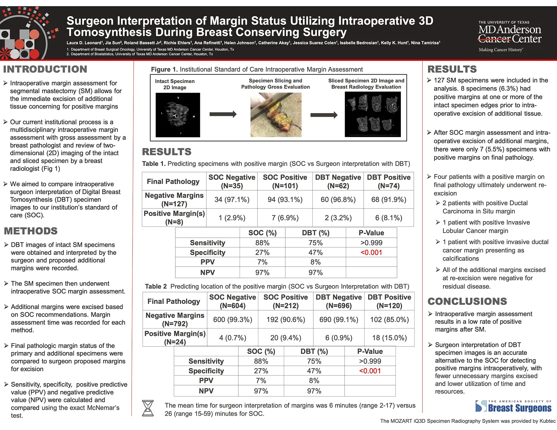
Nineteen of the 196 specimens (9.7 percent) had a positive margin before any additional shave margins were removed. Surgeon interpretation of the tomosynthesis images correctly flagged 16 of these 19 specimens, the same number identified by the standard multidisciplinary process, for matching sensitivity of 84 percent. Specificity told a different story: surgeons reading the 3D images achieved 49 percent specificity compared with 29 percent for the standard process (p<0.001), meaning fewer specimens without a true positive margin were flagged for additional excision.
When the analysis moved from the specimen level down to the 1,176 individual margins evaluated (38 of them positive), sensitivity was statistically similar across all three methods: 76 percent for the standard process, 68 percent for surgeon interpretation, and 74 percent for radiologist interpretation (p=0.61). Specificity again favored the tomosynthesis readings, at 87 percent for surgeons and 83 percent for radiologists versus 76 percent for the standard process (p<0.001). The two radiologists who reviewed the archived images independently showed fair agreement with each other (Cohen's kappa = 0.33), a reminder that 3D margin interpretation, like 2D interpretation before it, still carries some reader-to-reader variability.
The efficiency gain was substantial. Surgeons completed their tomosynthesis-based margin assessment in a median of 6 minutes (range 2 to 35), compared with a median of 33 minutes (range 15 to 88) for the full multidisciplinary review. After all additional shave margins guided by the standard process were excised, the final positive margin rate on permanent pathology was 4.6 percent (9 of 196 specimens), consistent with the low reoperation rates this institution has reported previously.
An earlier analysis of the first 127 specimens in this cohort, presented at the American Society of Breast Surgeons Annual Meeting in 2025, showed the same pattern: surgeon interpretation of tomosynthesis images achieved 47 percent specificity versus 27 percent for standard of care (p<0.001), with a mean interpretation time of 6 minutes versus 26 minutes.
Conclusions
Surgeon interpretation of intraoperative 3D tomosynthesis images offers sensitivity comparable to a multidisciplinary standard practice for detecting positive margins during breast-conserving surgery, with meaningfully higher specificity and a fraction of the time investment. The authors suggest this approach could reduce unnecessary shave margins without compromising oncologic safety, easing the resource burden that intraoperative margin assessment places on breast pathology and radiology teams.
Request your personal meeting or demo
Fill out the form and one of our exhibition managers with be in touch about scheduling your personal meeting or demo at our upcoming trade show.
For more news, views, & events, please visit our LinkedIn page
Click Here