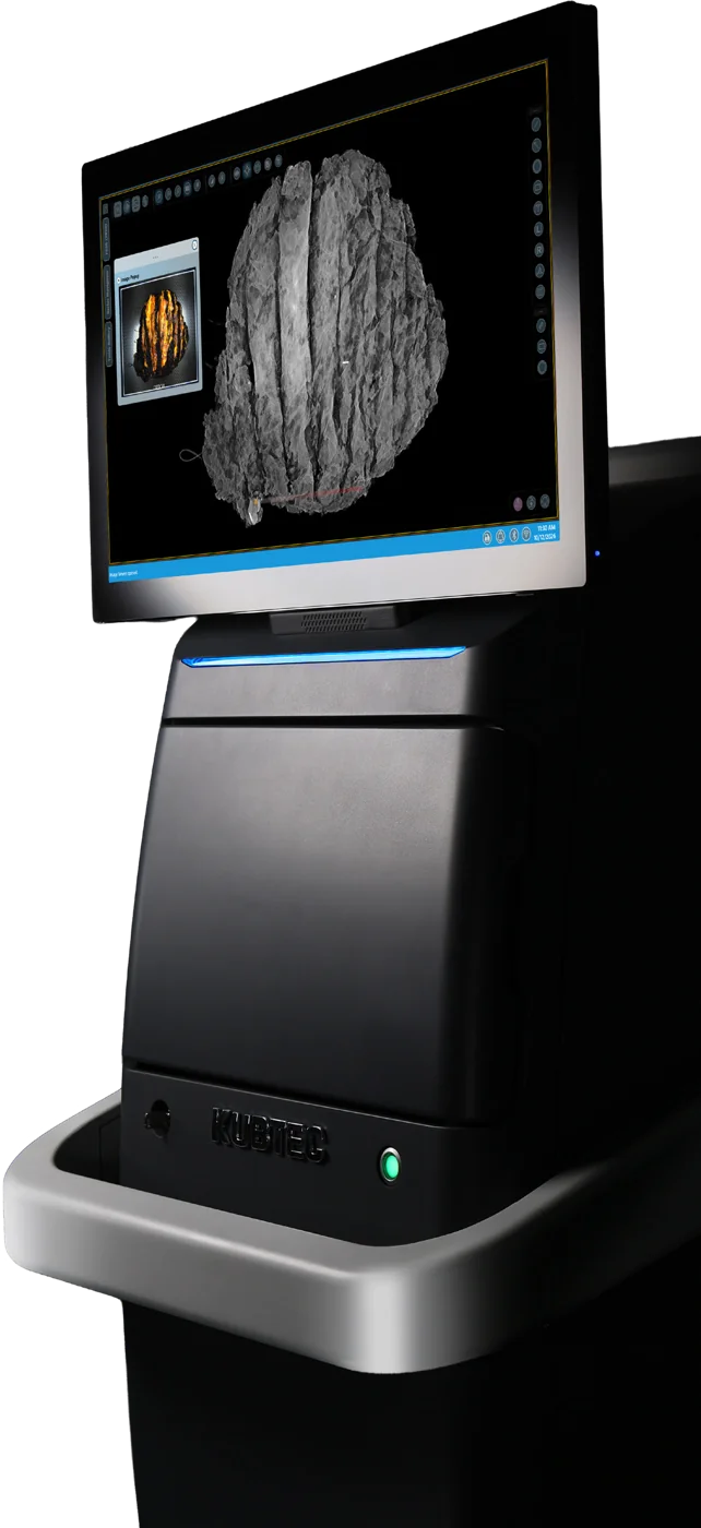
MOZART® Allegro 3D Specimen Radiography System

MOZART iQ Plus 3D Specimen Tomosynthesis System

2D was just the beginning.

The Future is 3D!

Contact us
MOZART iQ Plus 3D Specimen Tomosynthesis System
>50%
Fewer re-excisions vs Status Quo1
↓25%
Reduction in OR Time2
≥ 1 mm
Is emerging as the recommended minimum clear margin width for invasive breast cancer.3
apple slice
High quality 3D intraoperative margin assessment

Improved margin visualization, in three axes.

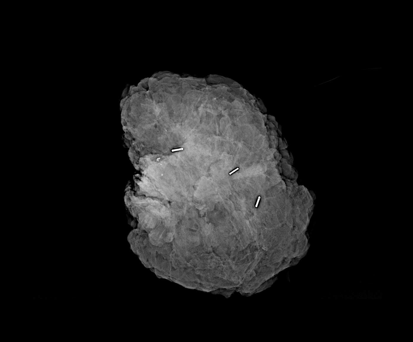
MOZART iQ Plus delivers high-quality 3D tomosynthesis imaging with 1 mm slices, allowing you to view the specimen slice by slice and see findings that may be hidden by overlapping tissue.Surgeons can assess the specimen and margins at excision, while PAs and Pathologists can use the same system during grossing to locate findings and guide tissue sampling.

3D Tomo with a larger imaging area

A 10" x 12" detector for larger specimens, like mastectomies, organs, and bones.

Choose a specimen to see it at true scale on each imaging area. Drag it to reposition, or set your own dimensions.

Choose a specimen
Or set your own dimensions (in)

Specimen:

MOZART iQ Plus 10" x 12" (25.4 x 30.5 cm) MOZART iQ 6" x 8" (15.2 x 20.3 cm) 2 in · grid lines every 1 in

Specimen sizes are illustrative examples, not clinical reference values. Imaging areas shown at true relative scale.

The System

Proprietary features designed around the way you operate.

3D Tomo with a Larger Imaging Area

A 10" x 12" detector for larger specimens, like mastectomies, organs, and bones. Compare imaging areas

Voice Control

Innovative incorporation of speech recognition technology allows you to operate the system without breaking scrub.

HD Optical Camera

Captures detailed optical images, enabling you to visually orient your specimen accurately, in real time.

The Image Blender™

Combines optical and X-ray images for the most comprehensive view of the specimen's anatomy.

Auto Magnification

Removes the guesswork with automatic, optimized image resolution.

X-Ray Image
Today's intraoperative standard, frozen sectioning, samples less than 5% of the specimen surface and takes 20 to 30 minutes while the patient remains under anesthesia.4 MOZART iQ Plus assesses the full specimen volumetrically, in the OR, with results before you close.
PEER-REVIEWED CLINICAL EVIDENCE

The Clinical Evidence That Sets MOZART Apart

MOZART 3D specimen tomosynthesis is backed by peer-reviewed clinical evidence, across hundreds of breast-conserving surgeries at leading institutions.

>50%
Decrease in re-excision rates vs. Status Quo1
↓25%
Reduction in OR Time2
≥ 1 mm
Is emerging as the recommended minimum clear margin width for invasive breast cancer.3
GammaPRO® + MOZART® iQ Plus

Integrated Gamma Detection

See your localization on the tablet or big screen, now with voice control. The GammaPRO® System works directly with KUBTEC Imaging Systems.

On the tablet or the big screen

Localization counts where the whole team can see them.

Voice control

Run localization hands-free, without breaking scrub.

One OR workflow

Localize with GammaPRO, then image the specimen on MOZART iQ Plus.

Explore GammaPro
BEYOND BREAST

Built for specimens of all sizes, from the OR to the gross room.

One system serving both Surgery and Pathology, from excision to gross examination.

Head & Neck
Breast
Thoracic / Lung
ENT
Bone and Soft tissue
GI
Colorectal, GYN
Placental

Smarter Software. Smoother Workflow.

One interface from login to archive. Start the case from the worklist, review the specimen in the image viewer, and send images where your team needs them, without leaving the system.

Modality Worklist

Start each case with patient and accession details already in place.

Image Viewer

Review, annotate, rotate, flip or invert images at the system.

PACS & Archive

Send images to PACS and keep an archive on the system.

Print & Export

Print or export images for the case record and pathology.