Breast specimen radiography helps identify lesions in tissue that is collected during breast-conserving surgery (BCS, also known as lumpectomy). The goal of the surgery is to remove all of the cancer while at the same time preserving as much healthy tissue as possible. To ensure that no tumor cells have been left behind, the surgical specimen needs to be X-rayed. Traditionally this has been performed using standard two-dimensional (2D) X-ray imaging. While beneficial, 2D imaging is extremely limited in what it actually enables the surgeon to see, as it doesn’t provide a full picture of the internal tissue of the surgical specimen. As a result, some cancer cells may be left behind, and consequently, many patients are called back for a second procedure.
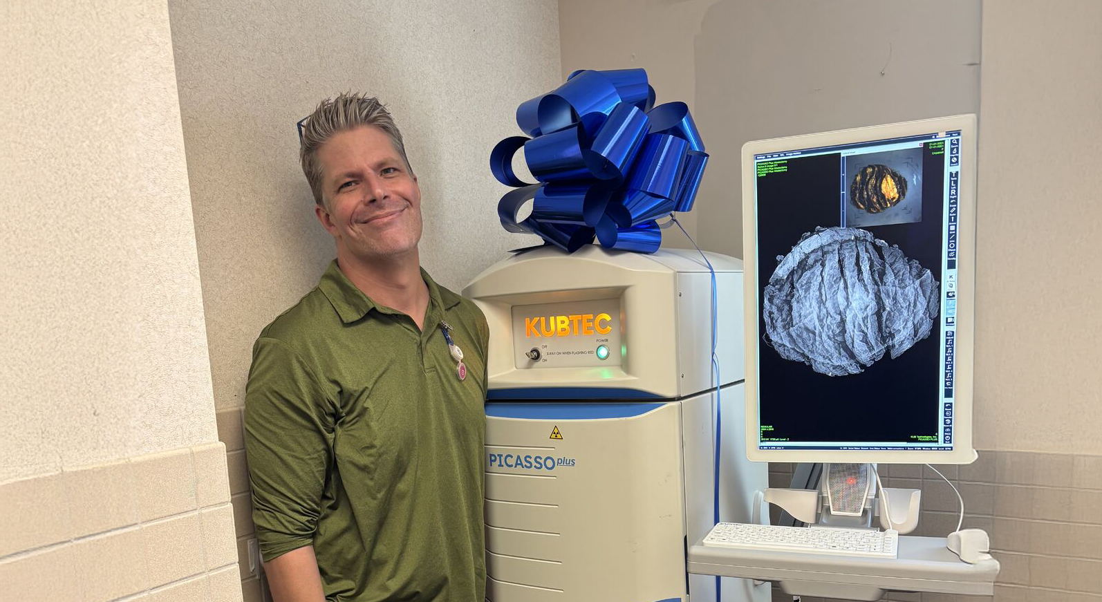
Now, a newer more advanced technology known as 3D tomosynthesis offers the solution to visualizing the internal tissue of the surgical specimen. Unlike its traditional 2D imaging counterpart, 3D tomosynthesis allows visualization of the breast specimen in three dimensions. This improves the surgeon’s ability to see the extent of the cancer and reduces the likelihood that cancer cells will be left behind. 3D tomosynthesis allows the surgeon to view the specimen in one-millimeter digital slices through the 3D image, providing the ability to see through overlapped breast tissue. This is especially helpful in viewing images of dense breast tissue by increasing the ability to visualize lesions and decreasing the number of second procedures, or re-excision.
In the following depiction of 3D tomosynthesis, each slice can be viewed independently and shows the top, middle, and bottom of the breast tissue, including the internal and otherwise invisible layers. Because each slice is unobscured by dense tissue above or below the area of interest, the surgeon or radiologist is able to clearly see margins around the lesion. Additionally, because the 3D system is present in the OR, the surgeon can determine, during the surgery, if more tissue needs to be removed or if the procedure is completed. This computes to a time savings and benefits for all concerned.

A traditional 2D specimen X-ray shows the three-dimensional breast anatomy on a single plane. Vertical perspective is lost, and overlapping breast tissue can hide breast cancers (known as a false negative) or make a normal spot appear to be abnormal (known as a false positive). This results in patient “callbacks” for more surgery and/or more imaging.
In the following depiction of 2D specimen imaging, there appears to be a clear margin (no cancer cells between the edge of the lesion and outer edge of breast tissue). However, during pathology examination of the specimen, the positive margin would be identified, and the patient would be scheduled for re-excision. This could have been avoided if 3D tomosynthesis was the technology used during this BCS. The surgeon would have been able to see the extent of the cancer, right there in the OR, and excise more tissue in the same procedure.

To try our interactive online demo of 3D tomosynthesis, please visit: Why 3D | KUBTEC (kubtec.com)
FREQUENTLY ASKED QUESTIONS
What is a surgical margin?
Wikipedia offers the following definition: “Surgical margin in a surgery report defines the visible margin or free edge of normal tissue seen by the surgeon with the naked eye. Surgical margin as read in a pathology report defines the histological measurement of normal or unaffected tissue surrounding the visible tumor under a microscope on a glass mounted histology section.”
Is there clinical evidence to support the use of 3D specimen radiographs?
Clinical evidence is available and highly supports the efficacy of 3D over 2D. The ability to see each slice of the specimen while in the operating room not only improves the patient experience but also increases the efficiency of the surgeon and the surgical staff.
What is breast conservation surgery?
According to the cancer.org site, breast conservation surgery (BCS) is an operation intended to remove breast cancer while still leaving as much normal tissue intact as possible. During this operation usually, some surrounding healthy tissue and some lymph nodes are also removed. Breast conversation surgery is also known as lumpectomy or partial mastectomy.
For more information about breast conservation surgery, visit these sites:
➡ Breast-Conserving Surgery | Northwestern Medicine
➡ Breast-Conserving Surgery | Cedars-Sinai
Request your personal meeting or demo
Fill out the form and one of our exhibition managers with be in touch about scheduling your personal meeting or demo at our upcoming trade show.
For more news, views, & events, please visit our LinkedIn page
Click Here