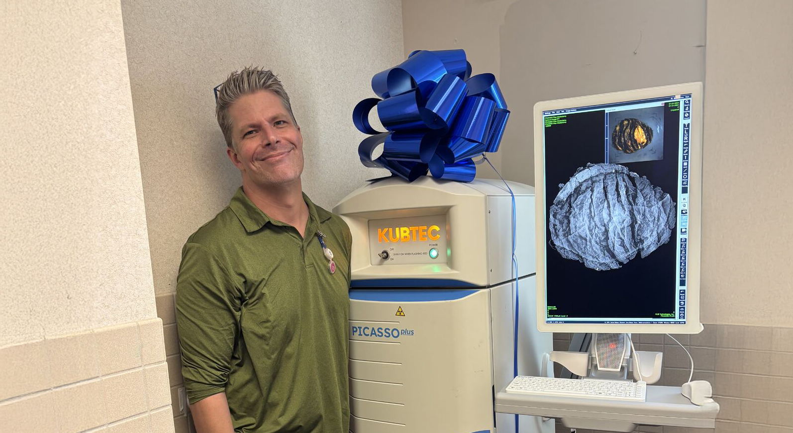
Three-dimensional tomosynthesis intraoperative analysis of screen detected breast malignancies reduces re-excision rates
A study from Royal Cornwall Hospital NHS Trust, Cornwall, England.
Background/Objective:
Breast-conserving surgery has generally been accepted as the treatment of choice for early invasive breast cancer. However, adequate local control depends on obtaining negative margins and receipt of radiation.
In 20-30% of patients with breast conserving surgery a second re-excision procedure is due to tumour-positive margins at final histopathology. These additional operations can produce considerable psychological, physical, and economic stress for patients and may delay use of recommended adjuvant therapies.
Margins re-excision rates are variable across the countries. Mean re-excision rate, across units in the UK is 17.2% .
Intraoperative specimen radiography, used to evaluate partial mastectomy specimens, ensures that the lesion is adequately removed.
Digital breast tomosynthesis (DBT)was approved by FDA in 2011 for breast cancer screening.
DBT creates a series of thin sliced, 2D images with angular rotation to provide z-axis resolution, resulting in improved lesion discernibility with increased cancer detection rates and fewer re-calls.
Tomosynthesis provides a specific depth of field, allowing surgeons to examine a specimen in one-millimetre increments and visualize the exact slice that best identifies the targeted tumour.
Methods:
Retrospective study comparing 2cohorts:
2D Cohort: 360screen-detected breast cancer 4/2015 to 3/2018
3D Cohort: 300screen-detected breast cancer 4/2018 to 3/2021
3D intraoperative system for all cases introduced at RCHT April 2018. Prior to this, the re-excision rate was stable at approximately 15% using established 2D intraoperative analysis.
• All patients had undergone preoperative digital mammogram and ultrasound.
• All malignancies were localised with ROLL(Radio-guided occult lesion localization).
• All wide local excision were performed by 5 oncoplastic breast consultants.
• Specimen radiography was performed intraoperatively using 2D X-ray or 3D tomosynthesis
• For both methods of assessment, specimen was placed in the device and auto-exposed without any compression of tissue.
• All specimens were:
• Marked with orienting sutures and clips according to local protocol
• Painted in theatre by the operating surgeon
• Examined by the same pathologists.
Results:
Statistical analysis was performed comparing 2D and 3D cohorts for:
• Patient demographics
• Histology
• Re-excision rates
Descriptive and comparative statistics were calculated for all collected data:
•3D/2DRR 0.59 (P:0.01),
•CI 0,3369-09068,
•Z 2.3473,
•P <0.02
Conclusions:
The use of intraoperative 3D specimen X-ray reduced the relative risk of re-excision rate by 41% (P=0.01) without any negative impact on other parameters.
References:
J. Mondani, H. Arabiyat, L . Kastora, M. Sulieman, I. Abbas, R. English, P. King,I. Brown, M. Barta, N. Jackson , P. Drew
The Mermaid Centre, Royal Cornwall Hospital, Truro Cornwall, UK, *Aberdeen Royal Infirmary , Breast Surgery Department Aberdeen, Scotland,UK
Request your personal meeting or demo
Fill out the form and one of our exhibition managers with be in touch about scheduling your personal meeting or demo at our upcoming trade show.
For more news, views, & events, please visit our LinkedIn page
Click Here


